A 59-year-old patient was referred to dental clinic for immediate dental implantation in the area of upper central incisors due to severe periapical lesions which not subjected to endodontic retreatment and periapical surgery. Removal of central incisors and periapical lesions, dental implant placement were performed under local anesthesia with sedation. During the surgery a significant bleeding was noted from a unilateral accessory canal (arrow). In our case, it exited at the palatal aspect of tooth 2.1; its foramen was of oval shape and measured 2.00 × 3.00 mm.
In case of that canal’s location it terms lateral incisive canal. Other similar terms can also be applied to describe this rare anatomical structure: accessory canal of the anterior maxilla, lateral incisor canal, and neurovascular variation in anterior palate.
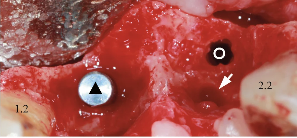
Panel clearly demonstrates the axial intraoperative view after bone drilling (circle) (preparing a hole for second implant); implant analog (which is used for correct positioning of implant) inserted into socket of tooth 1.1 is indicated by triangle. Lateral incisors marked by numbers 1.2 and 2.2.
About the Author
Ivan V. Nagorniak, Oral Surgeon, PhD, Private Dental Practice, Kyiv, Ukraine.
Back to Publications